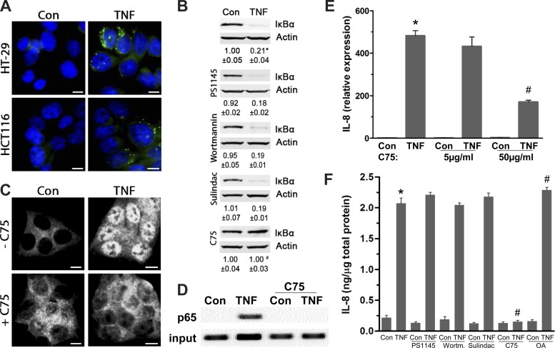
Fig. 4.
Increased LDs facilitate TNF-mediated IL-8 transcription by supporting NF-κB activity. A: HT-29 and HCT116 cells treated with TNF (6 h) were stained for LDs (BODIPY 493/503) (representative images from 3 independent experiments, scale bar = 5 μm). B: protein from HCT116 cells treated with TNF (0.5 h) with or without inhibitors for IKK-β (PS1145), PI3K (wortmannin), PGE2 synthesis (sulindac), or lipogenesis (C75) was immunoblotted for IκBα (n = 4, *P < 0.05, compared with Con, #P < 0.05, compared to TNF alone, ANOVA). C: immunofluorescent staining for p65, a NF-κB subunit (Alexa 594), in HCT116 cells treated with TNF (0.5 h) with and without C75 (representative images from 3 independent experiments, scale bar = 5 μm). D: chromatin immunoprecipitation (ChIP) assay of TNF-mediated p65 binding (0.5 h) to the IL-8 promoter with and without C75 treatment (50 μg/ml); input represents total chromatin levels before immunoprecipitation, indicating that equal amounts of samples were applied to ChIP followed by PCR. E: total RNA was extracted from HT-29 cells treated with TNF (2 h) in the presence of C75 (5 μg/ml and 50 μg/ml), and IL-8 transcription levels were determined with qPCR (n = 3, *P < 0.05, *compared with Con, #P < 0.05, compared with TNF alone). F: in HT-29 cells treated with TNF (4 h) with and without PS1145, wortmannin, sulindac, C75, or OA (24-h pretreatment), intracellular IL-8 protein levels were quantified by ELISA (n = 3, *P < 0.05, compared with Con, #P < 0.05, compared with TNF alone).

