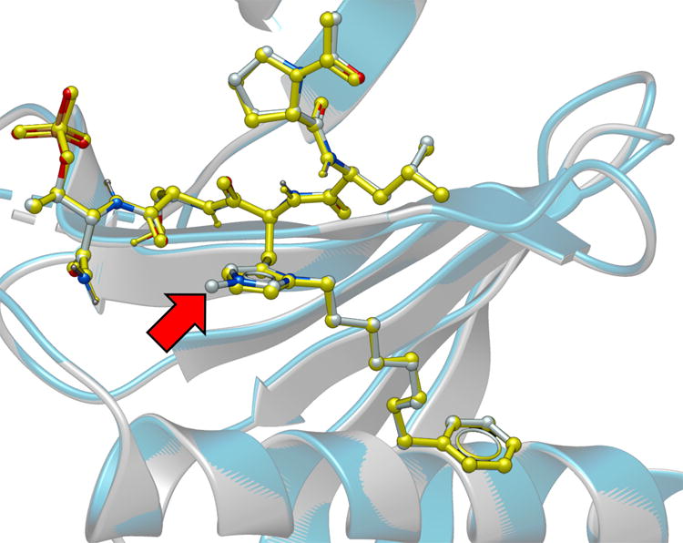
Figure 3.

Plk1 PBD co-crystal structure of 3m superimposed on the co-crystal structure of PBD-bound 2a (PDB 3RQ7), highlighting the high degree of correspondence. The 3m structure is rendered with the protein backbone shown as grey ribbon and the ligand colored according to element (carbon = grey, nitrogen = blue and oxygen = red). The 2a structure is rendered with the protein backbone shown as blue ribbon and the ligand colored yellow. The red arrow indicates the location of the resolved portion of the N(τ) substituent. Most of the alkyldiol substituent is unresolved.
