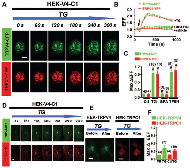
Figure 1.
Effect of Ca2+ store depletion on the translocation of TRPV4–C1 heteromeric channels to the plasma membrane, as measured by TIRFM. A through D, HEK cells were coexpressed with TRPV4-CFP plus TRPC1-YFP and were incubated with TG, 4 μmol/L. A and D, Time-series images of EFF for a representative single HEK cell (A) or a single vesicle (D). The bar indicates 5 μm in A and 1 μm in D. B, Time-series plot of EFF in representative cells for experiments similar to A. C, Summary data in experiments similar to A, showing the maximal change in EFF in response to TG or TPEN, 1 mmol/L. If necessary, BFA, 5 μmol/L, was introduced 30 minutes before TG application. E and F, HEK cells were expressed with either TRPV4-CFP or TRPC1-YFP alone. EFF images of a representative single cell (E) and summary data (F) before and after TG stimulation were shown. Controls in C and F were subjected to vehicle treatment (1% dimethyl sulfoxide). Data are given as the mean±SE (the number of experiments is labeled on top of the bars). #P<0.05 vs control, and *P<0.05 vs TG-treated cells without BFA.

