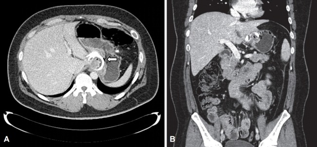
Fig. 2.

Abdominal and pelvic computed tomography for a routine health-check up in 2015. (A) Axial view, the gastric band partially eroded into the gastric lumen. The arrow shows that the gastric band caused gastric perforation. (B) Sagittal view, the eroded band is shown in the gastric lumen with gastric perforation.
