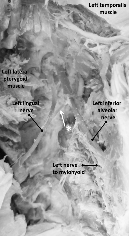
Figure 3. .

Photograph of dissected left infratemporal fossa. The left infratemporal fossa shows the mandible hemisected and turned over to expose the mandibular foramen with the left inferior alveolar nerve entering into the mandibular canal. The left lingual and inferior alveolar nerves show expected anatomy. A distinct nerve connects the lingual and inferior alveolar nerves, indicated by white asterisk and arrow.
