Figure 4. .
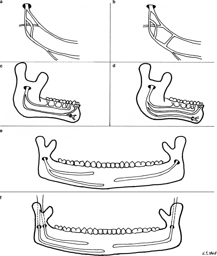
Summary diagram indicating variant branching patterns of the inferior alveolar nerve. (a) The inferior alveolar nerve can split high in the infratemporal fossa and then reunite to enter the mandibular canal as a single nerve. (b) During its course in the infratemporal fossa, the inferior alveolar nerve may be connected via small nerve branches to the lingual nerve. (c) A single inferior alveolar nerve entering the mandibular canal may course through the mandible as 2 distinct branches. These branches may be located within a single mandibular canal, or within 2 independent mandibular canals. (d) A single inferior alveolar nerve entering the mandibular canal may course through the mandible as 3 distinct branches. (e) Variations of the inferior alveolar nerve within the mandible may be unilateral. (f) A unilateral presentation of the inferior alveolar nerve may include a bifid nerve entering the mandibular canal as 2 distinct nerves, and continuing to course through the mandible as independent nerves.

