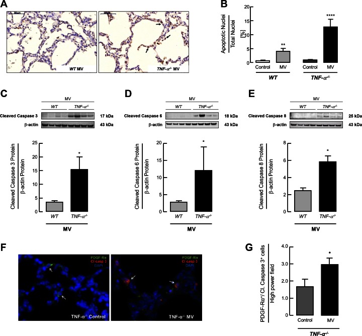
Fig. 2.
MV-O2 increases apoptosis in lungs of TNF-α−/− compared with WT mice. A: TUNEL staining of lung tissue sections, showing an increased number of apoptotic cells (black arrows) in the lungs of 6- t 7-day-old TNF-α−/− pups after 8 h of MV-O2 compared with ventilated WT mice. B: quantitative image analysis of TUNEL-positive cells indicating a significant increase in apoptosis in lungs of TNF-α−/− mice compared with WT littermates after 8 h of MV-O2. Significant difference between groups, **P < 0.01, ****P < 0.0001; n = 4–7/group. Immunoblot for protein expression showed a significant increase of pulmonary caspase-3 (C), caspase-6 (D), and caspase-8 (E) protein expression in newborn TNF-α−/− mice compared with WT littermates after 8 h of MV-O2. Significant difference between groups, *P < 0.05; n = 4/group. F: immunofluorescence image of lung tissue (×400, merged) showed increased dual staining for cleaved caspase-3 (red) and PDGF-Rα (green) in the lungs of 6- to 7-day-old TNF-α−/− mice after 8 h MV-O2 (panel at right) compared with unventilated controls (panel at left); white arrows indicate single (left) and dual (right) positive cells; nuclear counterstain with DAPI (blue). G: quantification of the images indicated an increase in dual positive cells per high-power field in 6- to 7-day-old TNF-α−/− mice after 8 h MV-O2. Significant difference between groups, *P < 0.05; n = 4/group; 10 high-power fields analyzed per mouse.

