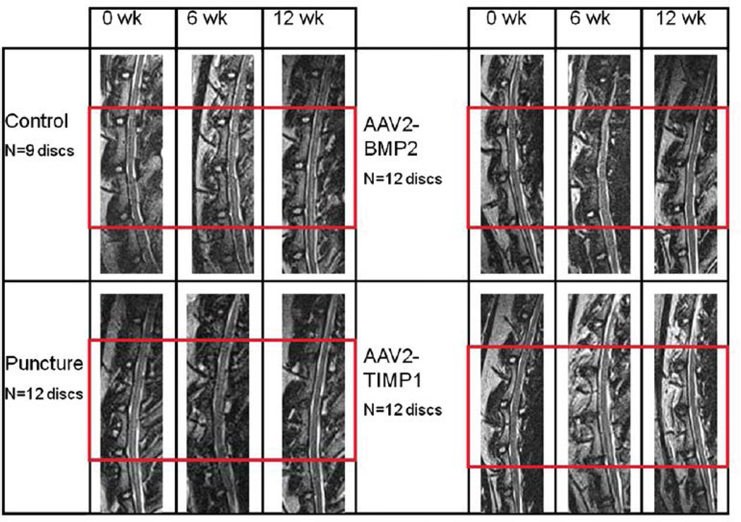
Fig. 5.
Lumbar spine magnetic resonance imaging (MRI) studies. Sample T2-weighted midsagittal lumbar MRIs of L1–L2 through L5–L6 at time points 0, 6, and 12 weeks. The treated discs (L2–L3, L3–L4, and L4–L5) are outlined by the red box. Sham surgery discs (not shown) look like nonpunctured control discs. AAV2-BMP2, adeno-associated virus serotype 2-bone morphogenetic protein 2; AAV2-TIMP1, adeno-associated virus serotype 2-tissue inhibitor of metalloproteinase 1.

