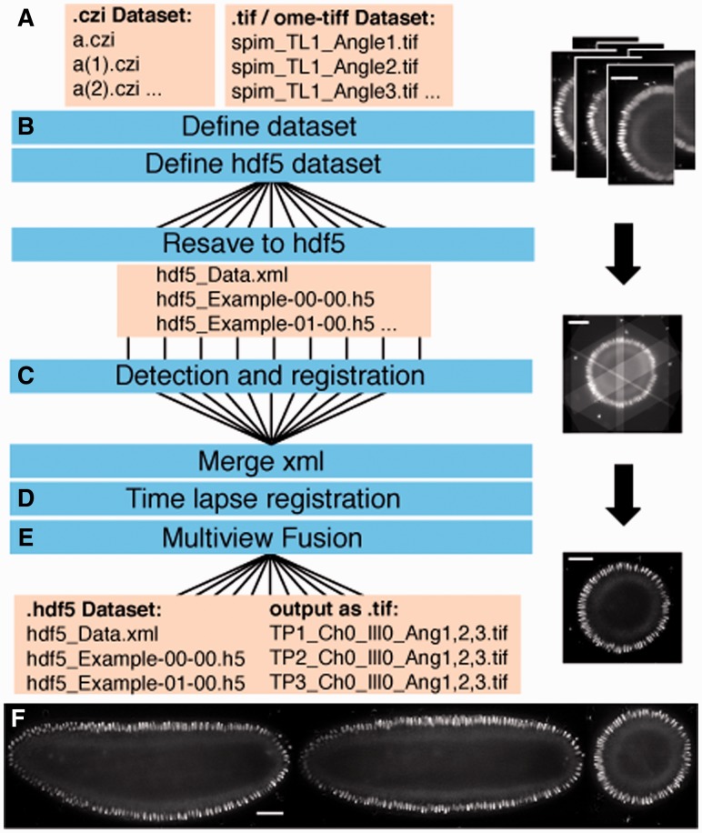
Fig. 1.
Automated workflow for multiview processing. Workflow for SPIM image processing (A–E) using parallelization (B, C and E). Shown on the right yz slices in the BigDataViewer of a Drosophila embryo expressing histone H2Av-mRFPruby raw (A) registered (C) and deconvolved (E). Results of deconvolution with xy , xz and xz slices through the fused volume of the same embryo (F). Scale bars represent 50 μm

