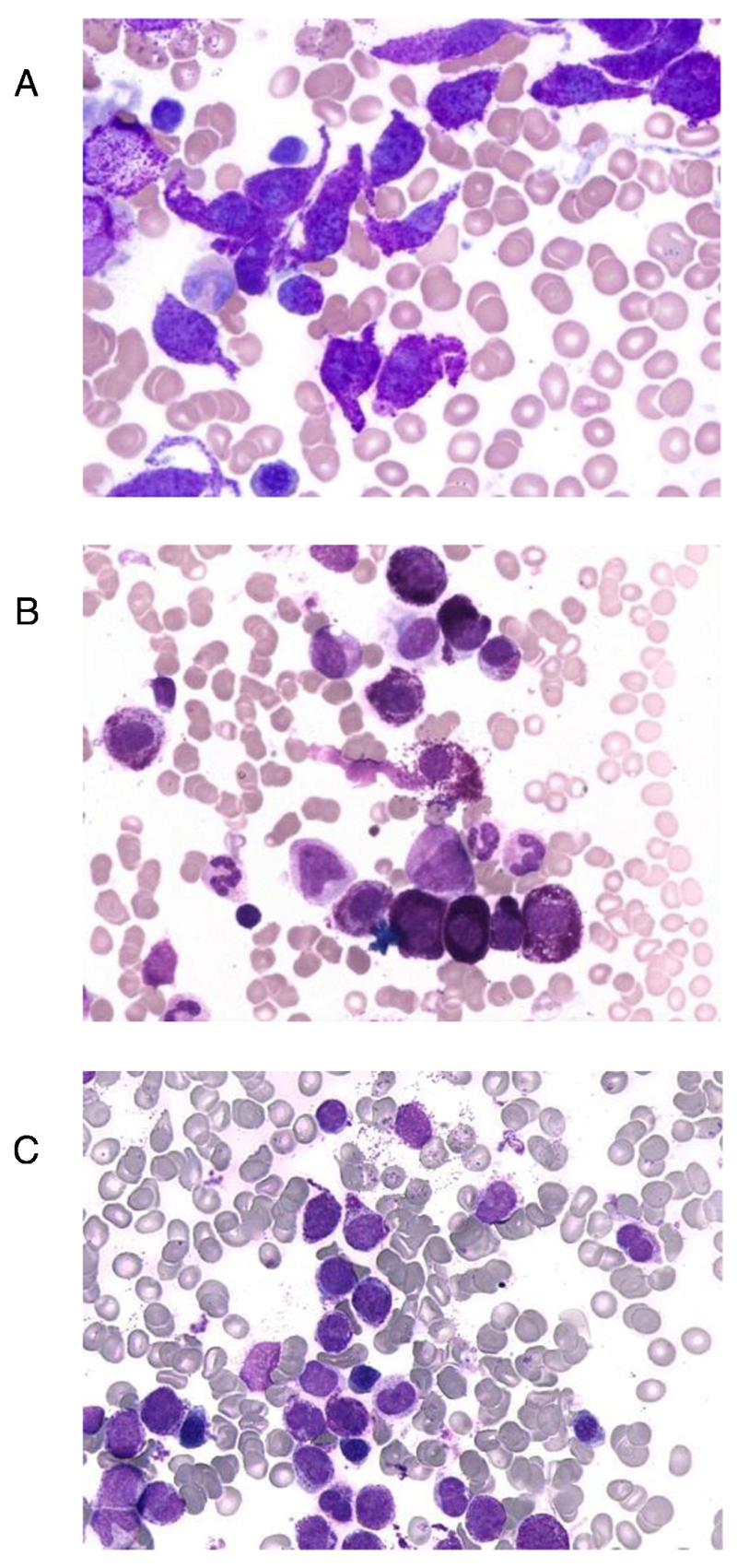
Figure 1.
Morphology of mast cells in chronic mast cell leukemia (MCL)
Bone marrow smears obtained from patients with chronic MCL (A, B) and acute MCL (C) were stained with Wright-Giemsa solution. In chronic MCL, mast cells are more mature cells, sometimes with a spindle-shaped (atypical) morphology (A) but often also with a mature morphology resembling normal tissue mast cells (B). By contrast, in patients with acute MCL, mast cells are immature and often represent metachromatically granulated blast cells (C).

