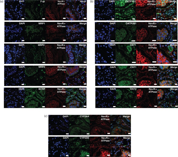
Figure 6.
Immunofluorescence imaging of selected transporters in fixed testicular tissue sections from a single HIV-1-infected, treated subject with representative images showing the localization pattern of (a) ABC transporters, (b) SLC transporters and (c) CYP450 metabolic enzymes. Tissue sections were stained with DNA dye, DAPI (blue) and examined by immunofluorescence using respective primary antibodies corresponding to our proteins of interest (green). Anti-Na+/K + ATPase-α (red) antibody was used as a marker for the plasma membrane. Cells were stained with Alexa Fluor-conjugated secondary antibodies 488/555 alone to verify the signal specificity of the primary antibodies (Figure S1a–c). Scale bar, 20 μm. This figure appears in colour in the online version of JAC and in black and white in the print version of JAC.

