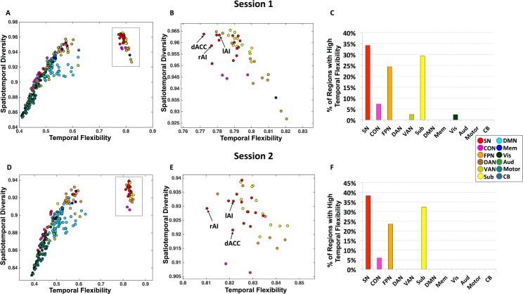
Fig 3. Brain regions with high temporal flexibility.
Panels A–C depict results from Session 1 data. (A) Joint profile of temporal flexibility and spatiotemporal diversity identifies a cluster of brain nodes with distinctly high temporal flexibility. (B) Detailed profile of brain nodes within the cluster with high temporal flexibility (inset from panel A). (C) Brain nodes with high temporal flexibility are primarily from the SN, subcortical regions, and FPN, with the highest percentage belonging to the SN. Panels D–F depict corresponding results from Session 2 data.

