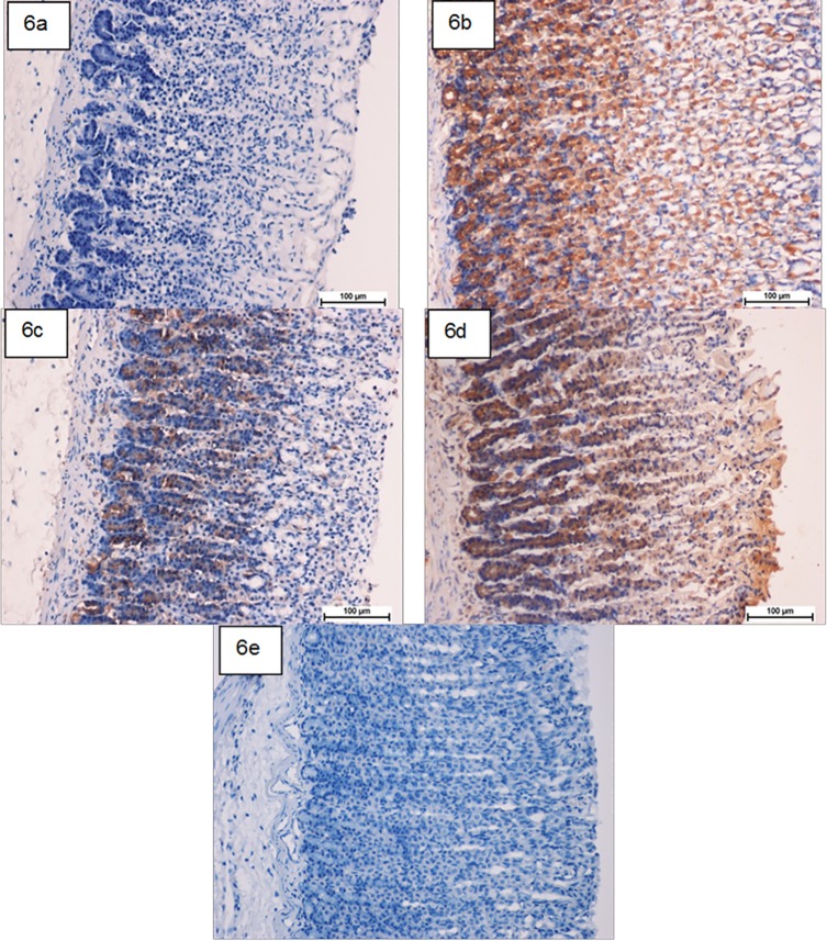
Fig 6. Photomicrographs showing the immunohistochemical analysis of HSP-70 protein.
(6a) ulcer control group, (6b) omeprazole group, (6c and 6d) the pre-treated groups with compound 1 at doses 50 and100 mg/kg, respectively. (6e) Rats in the normal control group showed intact gastric mucosa. The antigen site appears as a brown color (IHC: ×20).

