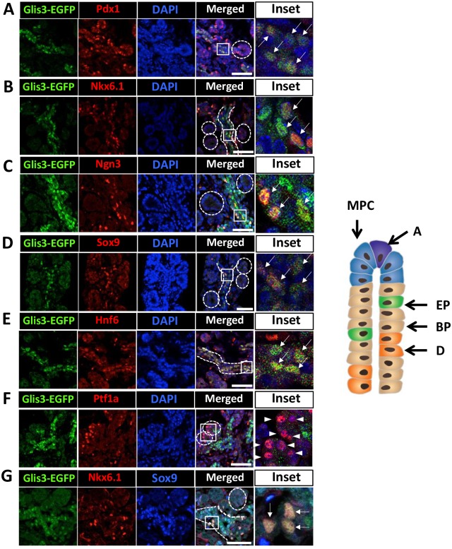
Fig 4. Glis3 protein expression at E15.5 of pancreas development.
E15.5 Glis3GFP/GFP pancreata were double-stained with antibodies against GFP (A-G) and Pdx1 (A), Nkx6.1 (B), Ngn3 (C), Sox9 (D), Hnf6 (E), or Ptf1a (M). DAPI stained and merged images are indicated. Boxed areas were enlarged and shown in the panels on the right. Arrows indicate Glis3+Pdx1+ (A), Glis3+Nkx6.1+ (B), Glis3+Ngn3+ (C), Glis3+Sox9+ (D) or Glis3+Hnf6+ (E) cells; arrow heads indicate Ptf1a single positive cells (F). Triple immunostaining of E15.5 Glis3GFP/GFP pancreata with anti-GFP, anti-Nkx6.1, and Sox9 (G). Arrows indicate triple positive cells (G, inset). Dashed circles indicate tip domains; dashed lines indicate trunk domains. Schematic view of tubule structure at E15.5 of embryonic pancreatic development is indicated on the right. DOI 10.6084/m9.figshare.3189172.

