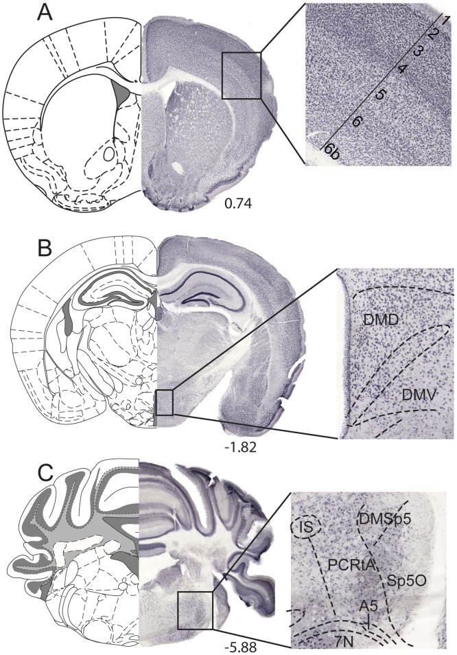
Fig 5. Abundant MFSD11 protein expression.
DAB-immunohistochemistry stained MFSD11 on 70μm wt floating brain sections. Overview images with specific regions magnified. (A) Staining pattern in cortex, (B) hypothalamic nuclei and the (C) brainstem. The schematic bregma regions were modified from The mouse brain [28].

