
Fig 6. Neuronal expression of MFSD5 and MFSD11 in the mouse brain.
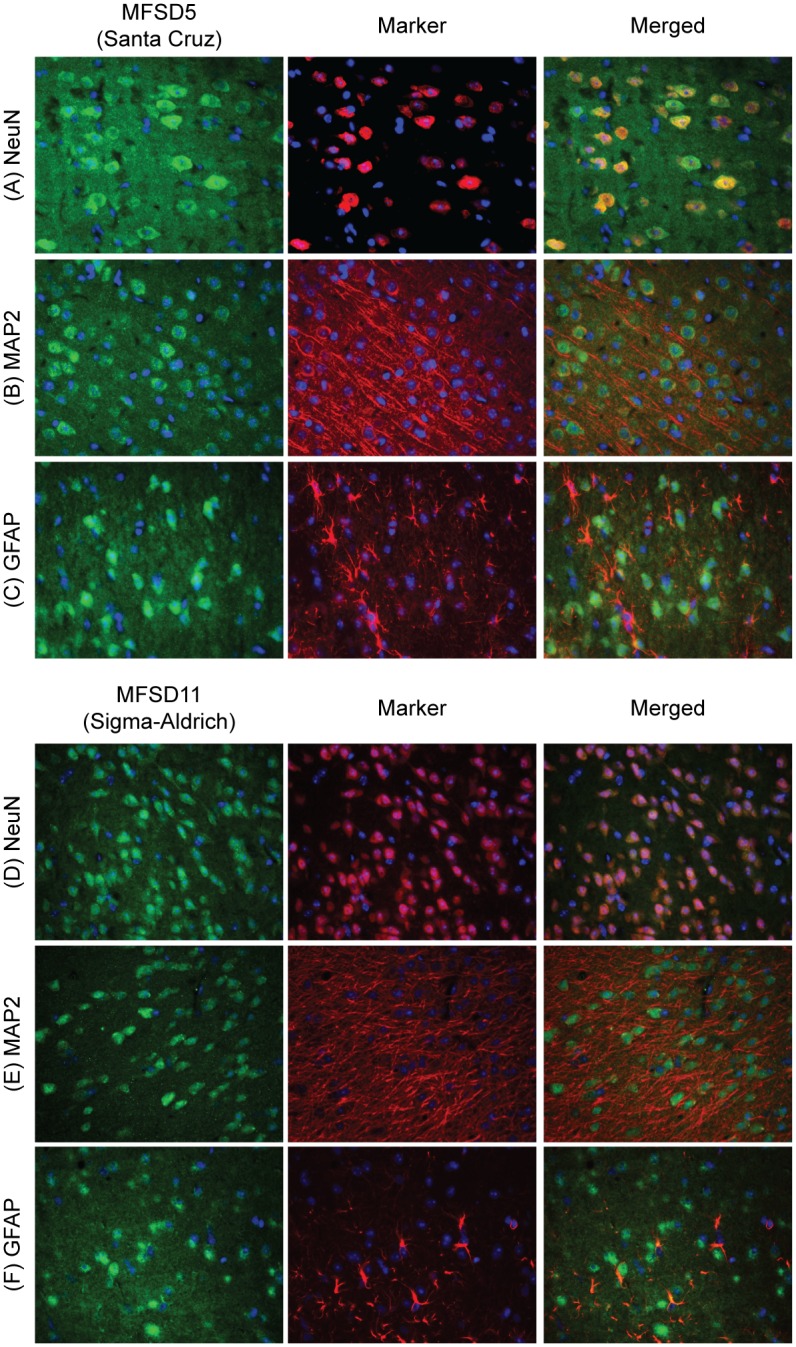
Double immunohistochemistry with fluorescent markers stained on 7μm wt mouse coronal paraffin sections. MFSD5 and MFSD11 were labelled in green, markers (NeuN, MAP2 and GFAP) in red and cell nuclei (DAPI) in blue. MFSD5 co-localized with the neural marker NeuN (A), but not with the neural dendritic marker MAP2 (B) or the astrocytic glial cell marker, GFAP (C). Also the MFSD11 antibody co-localized with NeuN (D), but not with MAP2 (E) or GFAP (F).