
Figure 5.
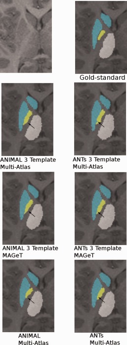
Using multiple templates for segmentation. Results for multi‐atlas and MAGeT Brain segmentations using three input templates. Black arrows pointing to region of visible improvement. In the ANIMAL case, the MAGeT Brain technique corrects the inaccurate segmentation on the medial wall of the globus pallidus. In the ANTs case, label ambiguity at the striatal‐pallidal edge is corrected. For both ANIMAL and ANTs, the full multi‐atlas‐based segmentation yields results most similar to the gold‐standard segmentation.