
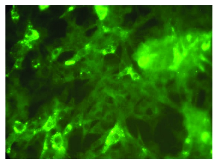
Figure 1. Showing the 50% end point dilution in the Rapid Fluorescent Focus Inhibition Test. Note approximately 50% BHK 21 cells in the microscopic field are infected as evidenced by presence of fluorescent foci representing rabies nucleoprotein (×400).