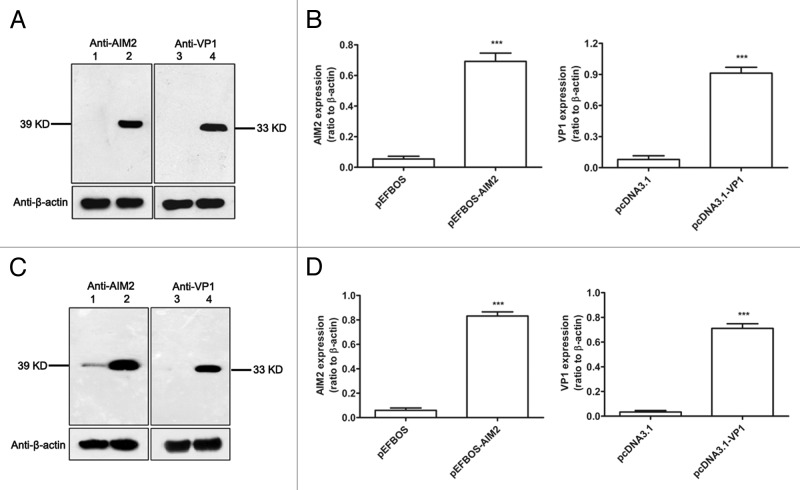
Figure 1. Expression of AIM2 and VP1 plasmids in vitro and in vivo. 293T cells were transfected with pAIM2, pVP1, or vector with lipofectamine for 48 h, and then cell lysates were subjected to western blot analysis using anti-AIM2, anti-VP1, or anti-β-actin antibody, respectively. (A) In vitro expression of AIM2 and VP1 plasmids. Lane 1: 293T cells transfected with pEFBOS vector; lane 2: 293T cells transfected with pEFBOS-AIM2; lane 3: 293T cells transfected with pcDNA3.1 vector; lane 4: 293T cells transfected with pcDNA3.1-VP1. (B) Quantification of pAIM2 and pVP1 expression by densitometry. Data are from one representative experiment of 3 performed and presented as the mean ± SD ***P < 0.001. BALB/c mice were intranasally immunized with chitosan-pAIM2 or chitosan-pVP1 vaccines, respectively, and intranasal mucosa biopsies were taken 3 d later for gene expression analysis. (C) Western blot analysis of AIM2, VP1, and β-actin expression for mucosal tissues from immunized mice. Lane 1: Mice immunized with pEFBOS vector; lane 2: Mice immunized with pEF-BOS-AIM2; lane 3: Mice immunized with pcDNA3.1 vector; lane 4: Mice immunized with pcDNA3.1-VP1. (D) Quantification of AIM2 and VP1 expression by densitometry in (C). Data are from one representative experiment of 3 performed and presented as the mean ± SD (n = 10). ***P < 0.001.

An official website of the United States government
Here's how you know
Official websites use .gov
A
.gov website belongs to an official
government organization in the United States.
Secure .gov websites use HTTPS
A lock (
) or https:// means you've safely
connected to the .gov website. Share sensitive
information only on official, secure websites.
