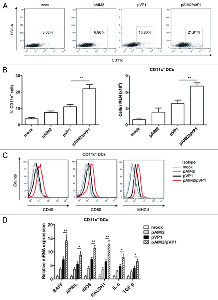
Figure 5. Increased DCs recruitment and IgA-inducing factors expression by co-immunization of chitosan-pAIM2 and chitosan-pVP1. Two weeks after the last immunization, MLN cells from mice in each immunization groups were analyzed. (A) The percentages of CD11c+ cells in MLN. One representative flow cytometry result was shown for per group. (B) Statistical analysis of the frequency and the absolute number of CD11c+ cells in MLN. (C) Representative expression profile of CD40+, CD80+, MHCII+ in DCs of MLN. (D) Real-time PCR analysis of BAFF, APRIL, iNOS, RALDH1, IL-6, and TGF-β relative to GAPDH for DCs cells from MLN. Data are from one representative experiment of 3 performed and presented as the mean ± SD (n = 10). *P < 0.05, **P < 0.01.

An official website of the United States government
Here's how you know
Official websites use .gov
A
.gov website belongs to an official
government organization in the United States.
Secure .gov websites use HTTPS
A lock (
) or https:// means you've safely
connected to the .gov website. Share sensitive
information only on official, secure websites.
