Fig. 5.
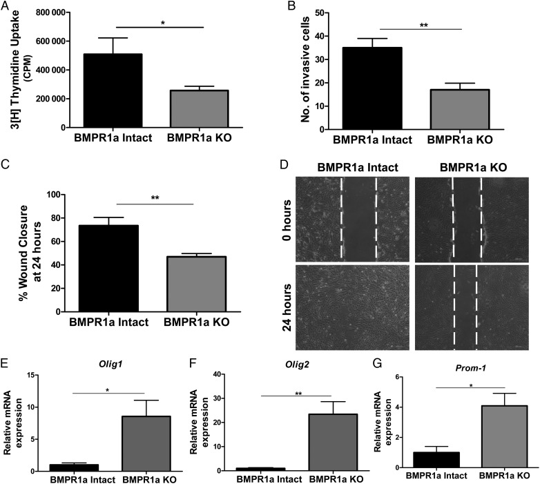
Abrogation of BMP signaling in transformed murine astrocytes inhibits proliferation, invasion, and migration and increases mRNA expression of stemness markers. (A)Tritiated thymidine incorporation assay shows that proliferation is inhibited in BMPR1a-KO astrocytes (gray bars) compared with BMPR1a-intact cells (black bars). Results are expressed as mean counts per minute (CPM) for quadruplicate samples at each time point for 3 cell lines per group. (B) In a 24-hour Matrigel transwell invasion assay, BMPR1a deletion results in a 50% reduction in the number of cells able to invade. Results are expressed as the mean number of cells able to invade with quadruplicate transwells and 3 cell lines per group. (C) Scratch assay was performed in 6-well plates with >90% confluent BMPR1a-intact and KO astrocytes. The BMPR1a-intact cells migrated into the wound, resulting in a smaller gap at 24 hours compared with the BMPR1a-KO cells. All cells were treated with mitomycin-C 2 hours prior to performing the scratch assay. (D) Representative images of BMPR1a-intact and BMPR1a-KO astrocytes at time of the original scratch (0 h) and at 24 hours (4x). (E,F,G)The mRNA expression levels of Olig-1 (E), Olig-2(F), and Prom-1 (G) were higher in BMPR1a-KO astrocytes compared with BMPR1a-intact astrocytes (n = 3 cell lines per group). A 2-tailed Student t test was performed to compare the mean mRNA expression. mRNA is normalized to Gapdh levels and relative to BMPR1a-intact expression. Error bars indicate SEM. *P < .05 **P < .01.

