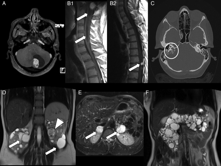
Fig. 1.
Diagnostic imaging in von Hippel-Lindau disease. (A) MR T1-weighted axial image with gadolinium showing a cystic cerebellar hemangioblastoma. (B1) MR T1-weighted sagittal image showing a brainstem hemangioblastoma with syringomyelia (arrows). (B2) MR T1-weighted sagittal image showing a thoracic spine hemangioblastoma (arrow). (C) CT scan showing a right endolymphatic sac tumor eroding the petrous bone (circle). (D) MR T2-weighted coronal image showing multiple bilateral renal cysts (arrows) and a left large clear cell carcinoma (arrowhead) characterized by inhomogeneous signal. (E) MR T2-weighted axial image showing large bilateral adrenal pheochromocytomas (arrows). (F) MR T2-weighted coronal image showing a polycystic pancreas.

