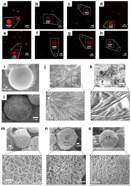
Figure 2.
Microscopy images of f-SHS@Guest: (a–h) in solution by CLSM, and in (i–o) the solid state by SEM. The guests corresponding to each image are (a) RhB; (b) Dox; (c) Cyt; (d) DsR; (e) DTR-3; (f) DTR-10; (g) DTR-70; (h) mCh. SEM images correspond to (i) f-SHS alone; (j) f-SHS@ Dox; (k) f-SHS@RhB; (l) lyophilized f-SHS; (m) f-SHS@DTR-3; (n) f-SHS@DTR-10; (o) f-SHS@DTR-70. The SEM images were drop-casted from a solution of 8ArG (0.303 mM, 121 mM KI, in 1X PBS, pH 7.4) and air-dried at 36 °C.

