Fig. S1.
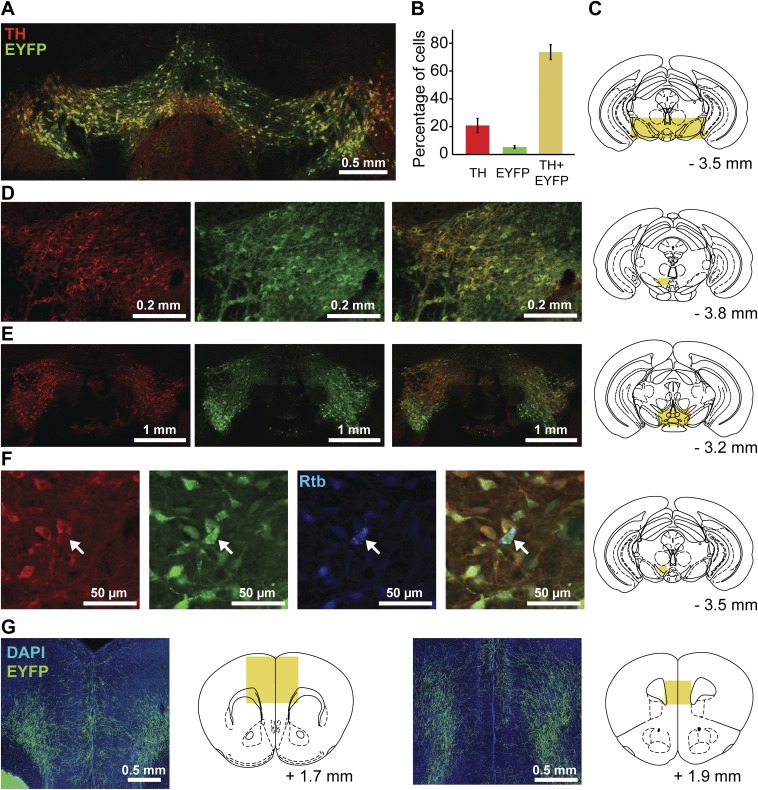
Expression of Cre-dependent EYFP in TH neurons and their mPFC projections. Expression of Cre enzyme in DA neurons was verified by injecting the AAV2-DIO-ChR2-EYFP construct in TH-Cre mice (n = 4) and performing immunostaining for TH in brain slices containing VTA. (A) Representative example of superimposed fluorescent channels (EYFP, green; TH, red). (B) Cell bodies expressing fluorescent markers (EYFP and/or Alexa 647) were counted (30 nonoverlapping images around the injection site from 16 slices, four slices from each mouse), and results were expressed as the percentage of total cells averaged across mice (n = 1,296). A small percentage of cells (5.3%) expressed the EYFP marker only, whereas the majority of cells (74%) were double-labeled. Limited viral diffusion from the injection site resulted in reduced recruitment of TH neurons from the lateral regions (A), with ∼21% of cells expressing the TH marker only. (C) Schematic drawing illustrating the location of the area displayed in A. (D and E) Additional histological examples from different mice showing TH staining (red), EYFP staining (green), overlay, and sampling location. (F) A different set of mice (n = 3) was injected with both AAV2-DIO-ChR2-EYFP in VTA and fluorescent retrograde beads (Rtb, blue) in mPFC, and then immunostained for TH. A representative example is shown. (Left to Right) TH, EYFP, Rtb, overlay, and sampling location. A total of 18 cell bodies were found expressing Rtb (25 nonoverlapping images from six slices, two from each mouse). Of these cell bodies, seven (39%) did not have either EYFP or TH fluorescence, three (17%) colocalized with TH staining, eight (44%) colocalized with both EYFP and TH, and none (0%) expressed Rtb and EYFP exclusively. (G) Examples showing a predominantly deep-layer distribution of EYFP-expressing axons in mPFC.

