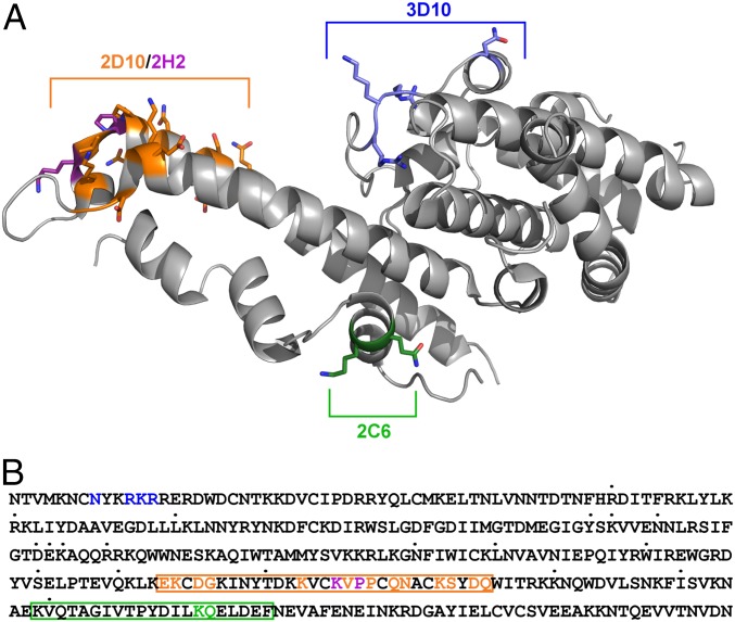
Fig. 6.
Epitopes of 2D10, 2H2, 2C6, and 3D10 mapped on PvDBP reveal that the epitopes are broadly conserved. (A) Epitopes are mapped on the surface of PvDBP with 2D10 in orange, 2H2 specific residues in purple, 3D10 in blue, and 2C6 in green. (B) Sequence of the Sal-1 DBP-II region with identified polymorphic sites indicated by dots above the individual residues. Structurally, mutationally, and HDX-MS (boxes)-identified epitopes are highlighted for 2D10 in orange, 2H2 in purple, for 2C6 in green, and for 3D10 in blue.

