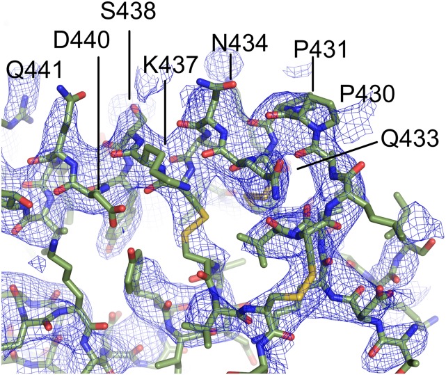
Fig. S1.
2Fo-Fc electron density contoured at 1σ around the epitope clearly identifies contact sites in DBP. The mAb is removed for clarity, and only DBP and associated electron density are shown. In this orientation, clear electron density for residues 433–441 that compose part of the epitope for 2D10 is observed.

