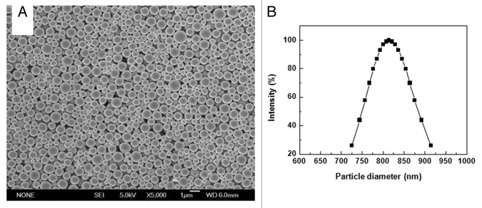
Figure 1. SEM photographs and size distribution of PLA microspheres. (A) Microsphere suspension was spotted on aluminum foil and dried naturally. Surface morphology of PLA microspheres was observed by SEM (B) Microspheres suspended in distilled water were examined by dynamic light scattering technology for size distribution.

An official website of the United States government
Here's how you know
Official websites use .gov
A
.gov website belongs to an official
government organization in the United States.
Secure .gov websites use HTTPS
A lock (
) or https:// means you've safely
connected to the .gov website. Share sensitive
information only on official, secure websites.
