
Figure 1. PET imaging studies of modulation of dopamine transporter function and expression of mGlu4 and mGlu5 in the brain of 6-OHDA lesioned rat during progressive degeneration.
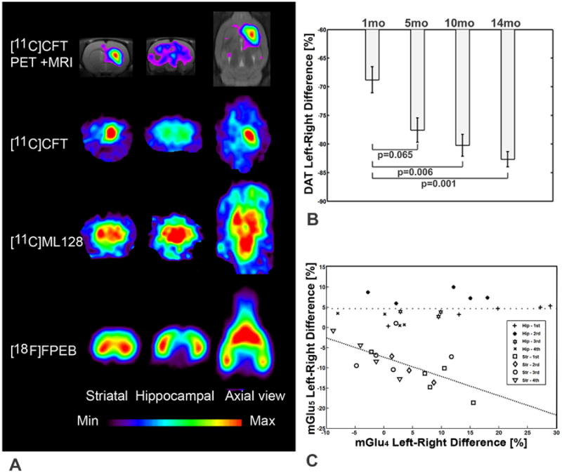
(A) Distribution of [11C]CFT ([11C]2β-carbomethoxy-3β-(4-fluorophenyl) on coronal (striatal and hippocampal level) and midstriatal axial slices shows abolished accumulation on the left (lesion) side. Distribution of [11C]ML128 (N-(4-chloro-3-[11C]methoxyphenyl)-2-picolinamide) on the same level as above shows enhanced accumulation on the left (lesion) side. Distribution of [18F]FPEB (3-fluoro-5-[(pyridin-3-yl)ethynyl] benzonitrile) on the same level as above shows decreased accumulation in the striatum and enhanced accumulation in the hippocampus on the left (lesion) side. (B) Exponential decline of dopamine transporter (DAT) binding of [11C]CFT in the striatum is an indication of progressive degeneration with the rate of 1.19±0.07 % per month. (C) Significant inverse correlation of mGlu4 and mGlu5 expression was observed in the lesioned striatum during progressive degeneration (ValuemGlu5 = (-0.478±0.477) × ValuemGlu4 - 7.34, r=0.647, p<0.01). mGlu4 expression declined in the hippocampus 102+/-82% while mGlu5 expression did not change during progressive degeneration.