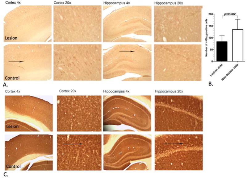
Figure 5. Representative pictures showing mGlu4 and mGlu5 immunostained rat brain.

Sections from the lesion and control side of the cortex and hippocampus fourteen months after the unilateral administration of 6-OHDA into the left nigra (magnifications ×4, ×20). (a) Arrows indicate mGlu4 expression area. At that time point mGlu4 expression had slightly decreased in the lesion side in the cortex and hippocampus of rat brain compared to non-lesion side. (B) Quantitative analysis using a paired t-test showed significant (p<0.002) decrease of the mGlu4 expressing cells on the lesion side of the hippocampus. (C) Immunostaining of mGlu5 expression did not show a significant difference between lesion and control side.
