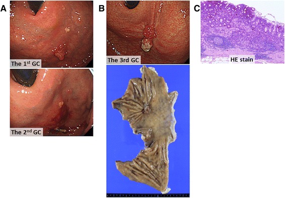
Fig. 1.

Findings of resected gastric cancers. a Gastroscopic images showing the first (upper) and second (lower) proximal gastric cancers. b Gastroscopic image of the third proximal gastric cancer (upper) and the macroscopic appearance of the resected proximal gastric specimen (lower). c Photomicrograph of a section from the resected gastric cancer showing well-differentiated tubular adenocarcinoma (hematoxylin and eosin stain, ×40)
