Fig. 1.

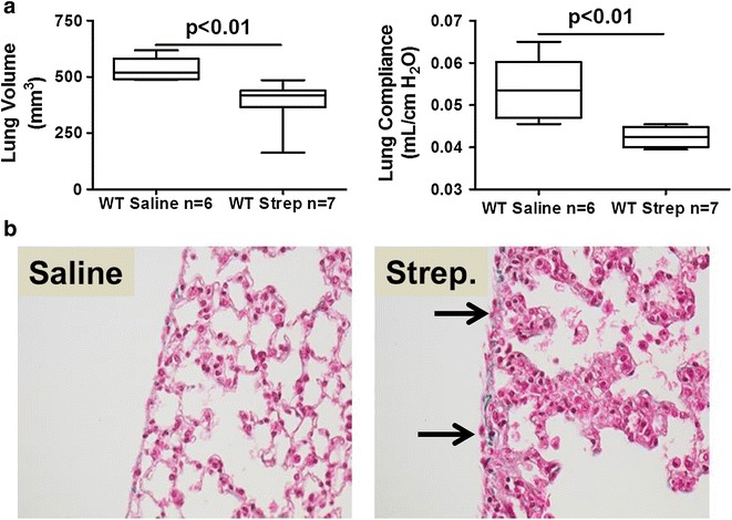
Intranasal S. pneumoniae causes pneumonia, lung dysfunction and mild pleural inflammation at survivable intranasal dosing. S. pneumoniae (3 × 10⁸CFU) was intranasally administered to WT mice and maintained for 7 days. a Lung volumes were measured by CT scan and pulmonary function (compliance) measured as described in the Methods. n = 6–7 mice/group. b Peripheral lung tissue sections from saline-challenged and S. pneumoniae infected mice collected at 7 days were stained with trichrome to show changes in lung architecture and collagen deposition (blue stain). S. pneumoniae infected mice exhibited thickened alveolar septae, mild pleural inflammation, reactive visceral mesothelial cells and mildly increased collagen deposition compared to saline controls. No pleural fluid or fibrinous strands at the pleural surface were seen. Solid arrows indicate reactive mesothelial cells. Images are 40× and are representative of the findings of 30 fields/slide and 6–7 mice/group
