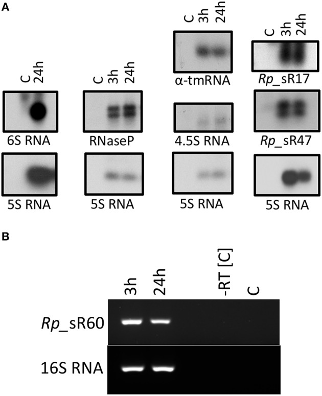
Figure 3.

(A) Northern blot analysis. A representative image of the Northern blots for candidate sRNAs is shown. Northern blot analysis was performed using strand-specific sRNA probes radiolabeled with [α-32P] UTP. All blots included RNA samples from control (uninfected HMECs) and those infected for 3 and 24 h with R. prowazekii. The blot for 6S RNA only included RNA isolated from HMECS that were either left uninfected or processed at 24 h post-infection. (B) Representative gel image showing the expression of sRNA Rp_sR60 during the infection of HMECs at 3 and 24 h post-infection.
