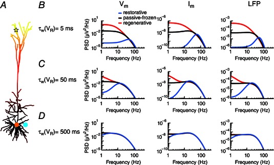
Figure 4. Activation time constants of active conductances determine the frequencies for which they affect the LFP .

A, apical white‐noise current input (yellow star) to the cortical pyramidal cell model with a single linearly increasing quasi‐active conductance (as in middle column of Fig. 3 B). B, the quasi‐active conductance is either restorative (blue), passive‐frozen (black) or regenerative (red), and the PSD is shown for the somatic membrane potential (left panel), the somatic transmembrane currents (middle panel) and the LFP (right panel) at a position close to the soma (cyan dot). The activation time constant of the quasi‐active conductance is C, as B, but with D, as B, but with
