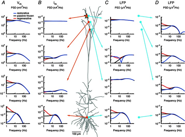
Figure 8. Membrane potential, transmembrane current and LFP for a pyramidal neuron with a single quasi‐active conductance .

White‐noise current input (yellow star) was applied to a cortical pyramidal cell model with a single quasi‐active conductance with increasing density with distance from soma, corresponding to the case in Figs 6 and 7. The quasi‐active conductance was restorative (blue), passive‐frozen (black) or regenerative (red). A, membrane potential PSD () is shown at the (intracellular) positions marked by the orange circles. B, as in A, but showing the transmembrane current PSD at the positions of the orange circles. C, as in A, but showing the LFP‐PSD for the extracellular positions marked by the cyan circles (20 μm from the corresponding cellular compartment marked in orange). D, as in C, but showing the LFP‐PSD for electrode positions (cyan circles) 600 μm from the corresponding cellular compartment marked in orange.
