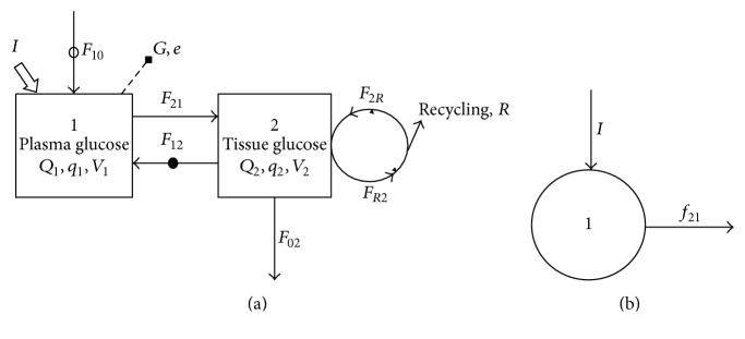
Figure 1.
Two-pool model for describing glucose kinetics (a) and the kinetic scheme for labeled glucose (b). The first pool represents total (labelled plus unlabelled) glucose in plasma, and second represents total glucose in tissues. Arrowed solid lines show flows, hollow arrow shows glucose application (infusion), a solid dot indicates a flow active only in the fasted state, a hollow dot indicates a flow active only in the fed state, and the broken line represents sampling. V i represents the volume of pool i, Q i and q i represent the quantity of total and labelled glucose in pool i, respectively, and F ij and f ij represent the flow of total and labelled glucose to pool i from pool j, respectively.

