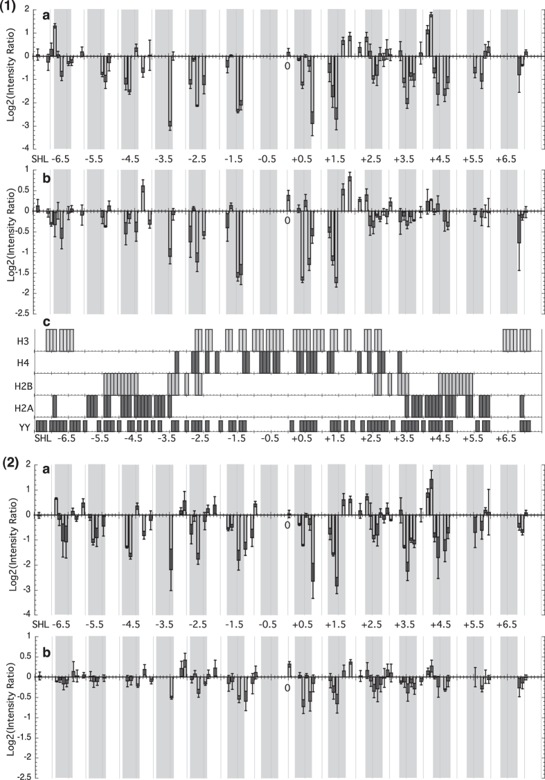
Figure 2.
Log2 of the intensity ratios of the peak height for Y-Y dimer peaks for reconstituted DNA compared with naked DNA with (1) 601 fragments and (a) histone (H3/H4)2 tetramer and H2A/H2B dimers, (b) only (H3/H4)2 tetramer. Changes in the ratio at each base step on both strands are shown. As additional information, (c) shows the DNA residues involved in the interface with H3, H4, H2A and H2B, and, just below, the location of pyrimidine-pyrimidine (YY) steps. (2) 601.2.4 fragments and (a) histone (H3/H4)2 tetramer and H2A/H2B dimers, (b) only (H3/H4)2 tetramer. Note that the y-ranges of panels (a) and (b) differ. Minor-groove inward facing regions observed in the nucleosome crystal structures are represented by grey boxes.

