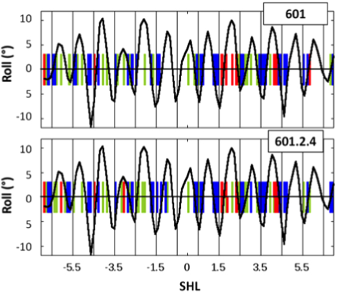
Figure 3.

Roll angles in X-ray structures of nucleosomes and changes in probabilities of Y-Y dimer formation upon histone octamer binding. The periodic variations of roll values along the DNA in nucleosomes are represented with a black line, using a natural smoothing spline approximation. These roll values were calculated and averaged on three X-ray structures of nucleosomes containing the 601 sequence (PDB codes 3LZ0, 3LZ1 and 3MVD). The rolls of the pyrimidine-pyrimidine steps that correspond to decreases and increases in probability of Y-Y dimer formation obtained by comparing DNA bound to the histone octamer and naked DNA are represented by vertical blue and red bars, respectively. The remaining pyrimidine-pyrimidine steps for which no change was observed are positioned by vertical green bars.
