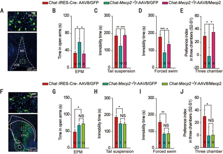
Figure 2.
Re-expression of Mecp2 in BF rather than CPu cholinergic neurons rescued behavioral deficits manifested in Chat-Mecp2−/y mice. (A) Immunohistochemical analysis showing the distribution of AAV8/Mecp2 in the BF of Chat-Mecp2−/y mouse brains 3 weeks after microinjection. (B) Anxiety-related behavior measured by the time in the open arms of the EPM. (C, D) Depression-related behavior measured by the immobility time that Chat-IRES-Cre and Chat-Mecp2−/y mice displayed in the tail suspension and forced swim tests. (E) Social behavior measured as the ratio of interaction time with a stranger mouse to interaction time with a familiar mouse in the three-chamber test. (F) Immunohistochemical analysis showing the distribution of AAV8/Mecp2 in the CPu of Chat-Mecp2−/y mouse brains 3 weeks after microinjection. (G-J) EPM, tail suspension, forced swim and three-chamber tests performed 3 weeks after injection of the AAV8/Mecp2 virus into the CPu. Error bars are means ± SEM. P-values were calculated by two-way ANOVA (genotype × trial) with Bonferroni's post hoc comparison. *P < 0.05, **P < 0.01, ***P < 0.001.

