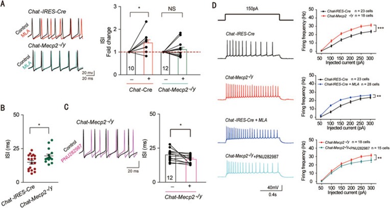
Figure 5.
Downregulated GABAergic excitability and upregulated glutamatergic excitability mediated by α7 signaling in the hippocampal CA1 region of Chat-Mecp2−/y mice. (A) Left: Representative APs of PV GABAergic interneurons in hippocampal CA1 in Chat-IRES-Cre mice (top) and Chat-Mecp2−/y mice (bottom) before (black) and after (color) MLA (10 nM) treatment. Right: Normalized ISI of hippocampal PV GABAergic interneurons before and after bath application of 10 nM MLA in both Chat-IRES-Cre and Chat-Mecp2−/y mice. P-values were calculated by paired t-test for each genotype. (B) Summary histogram of ISI of PV GABAergic interneurons in slices from Chat-IRES-Cre and Chat-Mecp2−/y mice (two-sided t-test). (C) Left: Representative APs of PV GABAergic interneurons in hippocampal CA1 in Chat-Mecp2−/y mice before (black) and after (color) PNU282987 (1 μM) treatment. Right: Plot of ISI before and after application of PNU282987 (1 μM). P-values were calculated by paired t-test. (D) Voltage responses to various current injection steps (1 s) in pyramidal neurons from Chat-IRES-Cre and Chat-Mecp2−/y mice at baseline or in the presence of MLA or PNU282987. Representative traces (left) and summary graph (right) show the firing frequency of pyramidal neurons from Chat-IRES-Cre and Chat-Mecp2−/y mice (two-way repeated measures ANOVA). Error bars are means ± SEM. *P < 0.05, ***P < 0.001.

