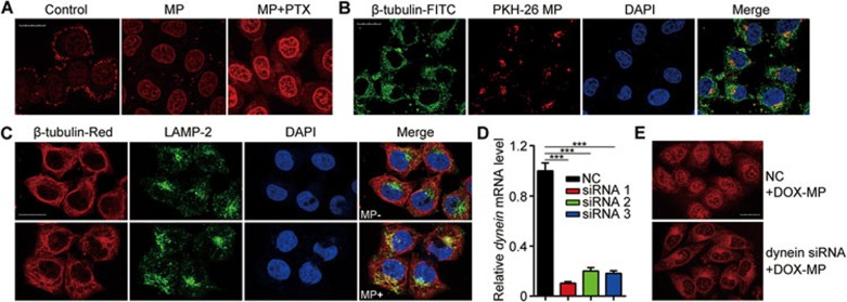
Figure 6.
Microtubules are involved in MP-mediated entry of drugs into the nucleus of TRCs. (A) PTX can enhance MP-induced drug entry into nuclei. MCF-7 cells with or without the 12-h MP pretreatment were treated with PTX (6 μg/ml) for 6 h. The cells were then treated with 1 μg/ml DOX for 4 h and observed under two-photon confocal microscope. (B) MPs co-localize with β -tubulin in MCF-7 TRCs. PKH26-labeled MPs were incubated with MCF-7 TRCs for 12 h. Then the cells were stained with FITC-labeled anti-β -tubulin antibody (green). DAPI was used to stain the cell nuclei (blue). (C) Much enhanced co-localization between LAMP-2 and β -tubulin in MP-treated MCF-7 TRCs. MCF-7 TRCs were incubated with MPs for 12 h. Then the cells were stained with fluorescence-labeled anti-LAMP-2 (green) and anti-β-tubulin (red) antibodies. DAPI was used to stain the cell nuclei (blue). (D) Efficiency of Dynein knockdown by siRNAs. Dynein siRNAs or control siRNA (NC) were transfected into MCF-7 TRCs. Twenty-four hours later, the expression of dynein was analyzed by real-time PCR. Error bars indicate mean ± SEM; n = 3 independent experiments. ***P < 0.001 (Student's t-test). (E) Dynein knockdown effectively blocked the entry of DOX into the nucleus. Dynein siRNA#1 or control siRNA were transfected into MCF-7 TRCs. Twenty-four hours later, MCF-7 TRCs were incubated with DOX-MPs for 12 h and then observed under two-photon confocal microscope. For all graphs, scale bars indicate 20 μm. See also Supplementary information, Figure S11.

