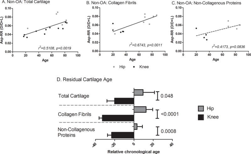
Figure 4. Calculated relative age of OA knee and OA hip cartilage matrix components.

To determine the biological relevance of the difference in racemization levels between OA hip and OA knee cartilage, we interpolated the biological age of an OA tissue specimen from the corresponding correlation of Asp racemization ratio with age for non-OA joints. Panels A–C show the age-related Asp-RRs for no-OA total cartilage (A), collagen fibrils (B) and non-collagenous proteins (C). The interpolated biological age was subtracted from the known chronological age of the sample to estimate the relative age due to disease. A positive relative age means the tissue is older than would be expected (lower protein turnover) while a negative value means the tissue is younger than expected (higher protein turnover). The Mann-Whitney test was used to test for significant differences in relative ages of cartilage by joint site. (D) OA knees were on average 30yrs ‘younger’ and hips 10yrs ‘older’ than non-OA joints. Of note, both OA knee collagen and non-collagenous components were ‘younger’ than the corresponding components of non-OA cartilage.
