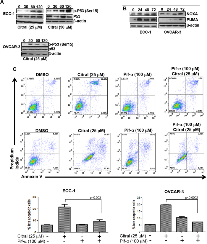
Figure 5. Citral-mediated activation of p53 results in apoptotic cell death.
A, ECC-1 and OVCAR-3 cells were treated with citral (concentration denoted below each western blot) for the designated time points. After treatment, cell lysates were probed for phosphorylation status of Serine-15 (Ser15) of p53 and for total p53. B, ECC-1 and OVCAR-3 cells were treated for the designated time points with citral (25 μM). Cell lysates were monitored for expression of p53-responsive genes PUMA and NOXA in both cell lines. C, ECC-1 and OVCAR-3 cells were treated with citral in the presence or absence of the p53 inhibitor pifithrin-α (Pif-α). After incubation for 24 h, apoptosis in the cells was determined by annexin V-FITC and propidium iodide staining. Cells were monitored using flow cytometry. Dot plots in the upper and lower panels are representative data obtained from ECC-1 and OVCAR-3 cells, respectively. The bar charts at the bottom show percentage of apoptotic cells after treatment from three independent experiments.

