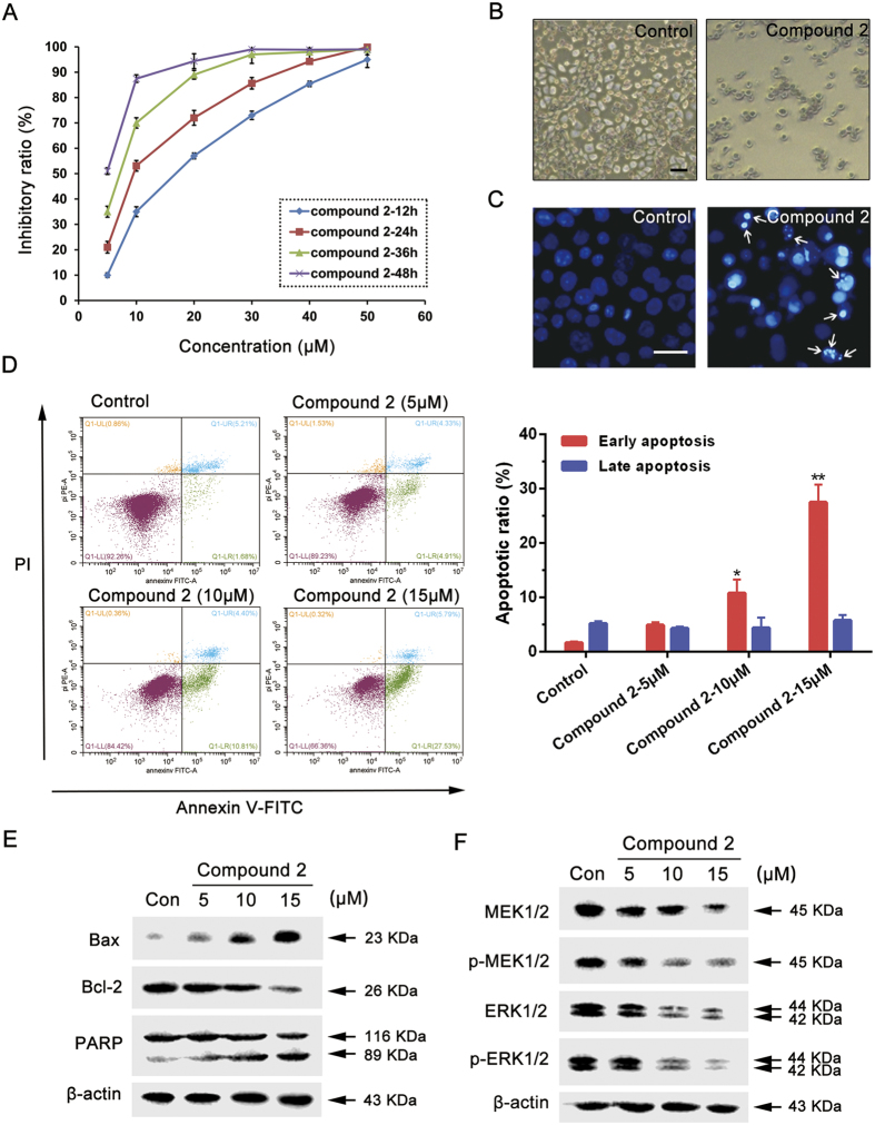
Figure 7. Compound 2 induces apoptosis in MCF-7 cells via targeting MEK/ERK pathway.
(A) Effect of compound 2 on MCF-7 cell viability. The cells were cultured for 24 h, and then incubated with different concentrations of compound 2 for 12, 24, 36 and 48 h. The viability was determined by the MTT assay. The data are presented as the mean ± SD of the results from three independent experiments. (B) The cells were treated with compound 2 (10 μM) for 24 h, then observed by phase contrast microscope. Scale bar = 100 μm. (C) The cells were treated with compound 2 (10 μM) for 24 h then stained with Hoechst 33258 and observed by fluorescence microscope. Arrows indicate the apoptotic bodies. Scale bar = 50 μm. (D) The cells were treated with different concentrations of compound 2 for 24 h, then revealed by Annexin-V/PI double staining using flow cytometry analysis. *Compared with control, p < 0.05, **Compared with control, p < 0.01. (E) The cells were incubated with different concentrations of compound 2 for 24 h, then the expression levels of Bax, Bcl-2 and PARP were detected by western blot analysis. Each lane was loaded with 30 μg of whole cell extracts, β-actin was used as a loading control. (F) The cells were incubated with different concentrations of compound 2 for 24 h, then the expression levels of MEK1/2, p-MEK1/2, ERK1/2 and p-ERK1/2 were detected by western blot analysis. Each lane was loaded with 30 μg of whole cell extracts, β-actin was used as a loading control.

