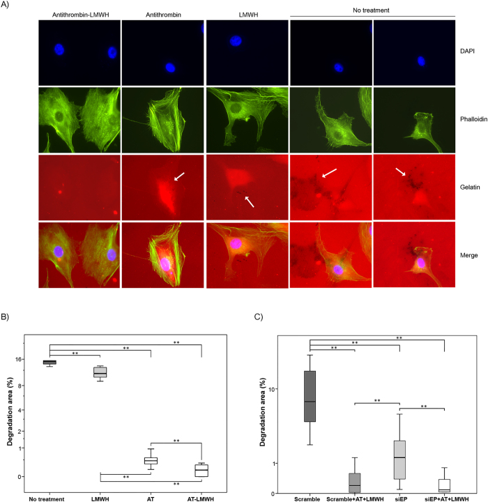
Figure 6. Effects of antithrombin and heparin on invadopodia degradation of gelatin coated coverslips.
(A) Representative micrographs of U-87 MG cells on a rhodamine-gelatin matrix. Cells were incubated without treatment or with low molecular weight heparin (LMWH), antithrombin (AT) and antithrombin activated by LMWH (AT-LMWH). Invadopodia were identified by colocalization of actin punctae over or close to areas of degraded matrix, which are indicated with a white arrow. (B) Quantification of invadopodia-mediated gelatin degradation of cells under the different treatments. (C) Quantification of invadopodia-mediated gelatin degradation of cells transfected with a scrambled siRNA or the specific silencer for TMPRSS15 gen (SiEP) with or without treatment with AT in combination with LMWH. Each condition was assayed in triplicate, and up to twenty-five different images were processed for each condition; *p < 0.05; **p < 0.01. Images were acquired with a Nikon 90i microscope at 60×, analyzed with Fiji software to calculate gelatin degradation and statistical analysis was carried out with a Mann-Whitney U test.

