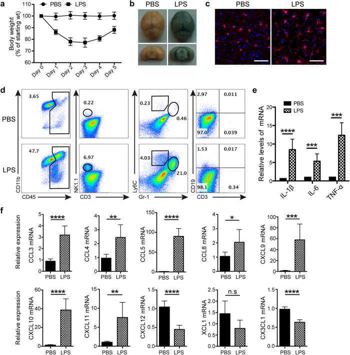
Figure 1. NK cells infiltrate into the brain after LPS treatment.
(a) C57BL/6 mice were injected i.p. of LPS (2 mg/kg/day) diluted in PBS for 5 days, and the body weight was measured (n = 5~6 at indicated time). Mice treated only with PBS acted as the control (n = 6). (b) Mice-treated with PBS or LPS for 3 days received i.v. 0.1 ml of 4% Evans Blue perfusion. One hour later, mice were killed and the whole brain as well as coronal section were prepared to evaluate Evans Blue extravasation. (c) Following administration of PBS or LPS for 3 days, morphology of microglia in the brain was analyzed by immunofluorescence staining with Iba-1 (red) and DAPI (blue). Bars, 100 μm. (d) After 3 days of PBS or LPS treatment, single cell suspensions from the whole brain were prepared and analyzed by flow cytometry. CD45+ leukocytes were gated and analyzed for identification of cell types. (e,f) Twelve hours after LPS or PBS treatment, mRNA were extracted from brain of mice (n = 6 per group). qPCR was performed to detect the expression of proinflammatory cytokines and chemokines. *P < 0.05, **P < 0.01, ***P < 0.001, ****P < 0.0001, unpaired Student’s t test. Means ± SD are shown. (b–d) Data shown are representative of 4 mice per group. All data in this figure are representative of 3 independent experiments.

