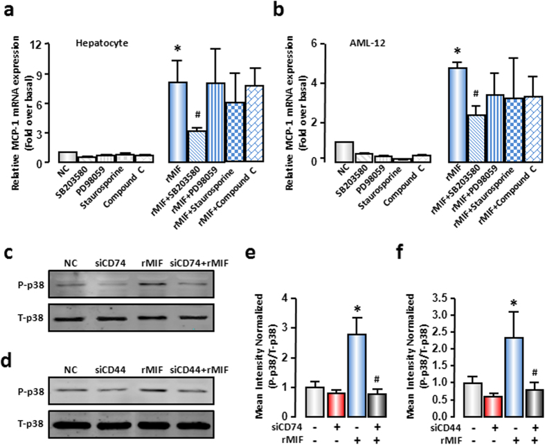
Figure 5. MIF promoted MCP-1 expression through p38 MAPK.
(a) Mouse primary hepatocytes or (b) AML-12 cells were pre-treated with SB203580 (10 μM), PD98059 (10 μM), staurosporine (10 nM), compound C (10 μM) for 1 hour, and followed by 100 ng/mL rMIF treatment for another 6 hours. MCP-1 mRNA expression was evaluated by real-time RT-PCR. (c,d) AML-12 cells were transfected with SCR siRNAs, CD74 siRNAs or CD44 siRNAs. After 48 hours, cells were treated with rMIF, after another 6 hours, cells were collected. Total p38 and phosphor-p38 levels were evaluated by Western blot analysis. Typical autoradiograms were shown. (e,f) The quantitative assay of Western blot was executed by Odyssey® software. All results were confirmed in three independent experiments at least. *P < 0.05 vs untreated control cells. #P < 0.05 vs rMIF treated cells alone.

