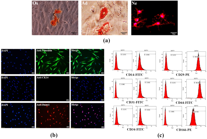
Figure 2. Characterization of hPDLSCs and examination of DNMT1 expression in hPDLSCs.
Immunocytochemical analysis showed that isolated hPDLSCs were negative for CK14 and positive for vimentin. Additionally, DNMT1 was localized to the nuclei and abundantly expressed in hPDLSCs (a). Flow cytometric analysis showed that cultured hPDLSCs were negative for CD14 (0.90%), CD31 (0.40%) and CD34 (0.54%) but positive for CD29 (99.92%), CD44 (98.45%) and CD166 (61.92%). Mouse IgG isotype control antibodies conjugated to FITC or PE were used as negative controls (b). After being separately cultured in osteogenic (Os) or adipogenic (Ad) medium for 21 days, mineralized nodules were stained with alizarin red solution and oil droplets were stained with oil red O solution. hPDLSCs that were cultured in neurogenic (Ne) media for 2 hours formed axon-like structures (c).

