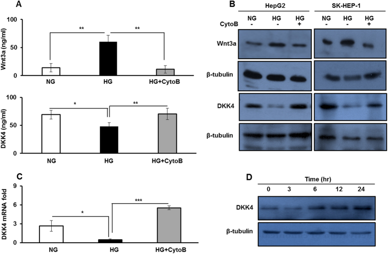
Figure 2. High glucose increases Wnt3a level and suppresses the expression of its antagonist DKK4.
(A) ELISA measurements of Wnt3a and DKK4 secretory proteins in culture media collected after 16 hr from HepG2 cells in NG, HG and HG + CytoB. (B) HepG2 and SK-HEP-1 cells were cultured in NG, HG and HG + CytoB for 16 hr. Whole cell lysates were subjected to western blotting and levels of Wnt3a and DKK4 proteins were detected. (C) HepG2 cells were cultured in NG, HG and HG + CytoB for 16 hr. Total RNA was isolated and cDNA was prepared to determine relative mRNA fold expression of DKK4 by quantitative real-time RT-PCR. (D) HepG2 cells were cultured in HG for 16 hr and then allowed to grow in medium without glucose for indicated time course. Whole cell lysates were prepared for detection of DKK4 protein by western blotting. All the bar graphs represent the mean ± SD of an experiment done in triplicate (*P < 0.05, **P < 0.001, ***P < 0.0001). Cropped blots are used in the main figure and full length blots are included in Supplementary Figure 7.

