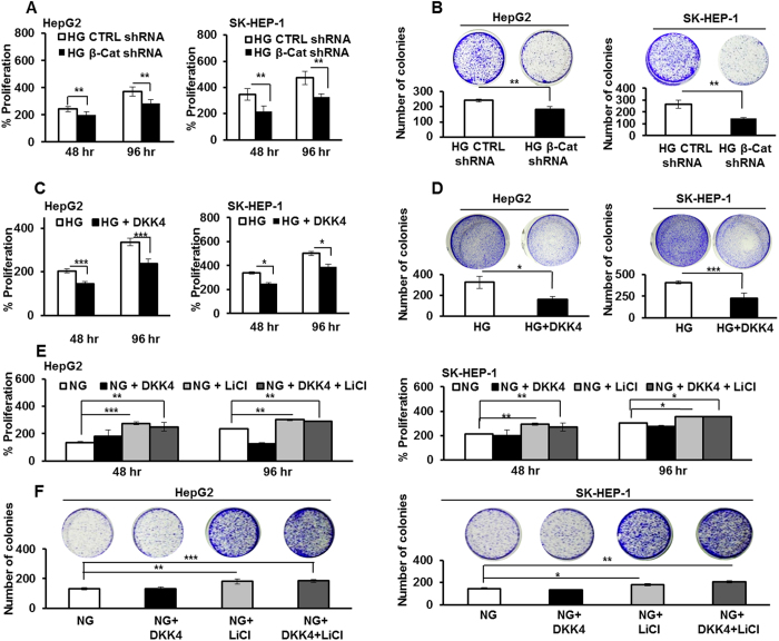
Figure 4. Glucose induced proliferation is dependent on β-catenin expression.
(A) HepG2 and SK-HEP-1 cells were cultured in HG and transfected with β-catenin shRNA or control shRNA and percentage proliferation was determined by MTT assay. (B) HCC cells were transfected with β-catenin shRNA or control shRNA. Post 48 hr of transfection cells were cultured for additional 21 days. Thereafter, colonies were visualized by crystal violet stain and counted. (C) HCC cells were cultured in HG and HG + DKK4 protein in culture medium for 48 hr and 96 hr. MTT assay was performed and percentage proliferation was determined. (D) HCC cells were cultured in HG and HG + DKK4 recombinant protein in culture medium for 48 hr and cells were cultured for additional 21 days. Thereafter, colonies were visualized by crystal violet stain and counted. (E) HepG2 and SK-HEP-1 cells were cultured in NG, NG + LiCl, NG + DKK4 and NG + LiCl + DKK4 protein, for 48 hr and 96 hr. Percentage proliferation was determined by MTT assay. (F) Colony formation assay in HCC cells cultured in NG, NG + LiCl or NG + DKK4 or NG + LiCl + DKK4 protein, for 48 hr and cells were cultured for additional 21 days. Thereafter, colonies were visualized by crystal violet stain and counted. All the bar graphs represent the mean ± SD of an experiment done in triplicate (*P < 0.05, **P < 0.001, ***P < 0.0001).

