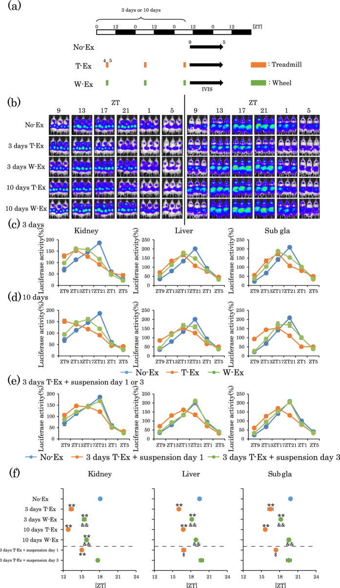
Figure 1. Treadmill or wheel-running exercise during the inactive period produces phase advance of mouse peripheral clocks.
(a) Experimental schedule. The white and black bars indicate environmental 12 h light and dark conditions. The orange short bar indicates forced exercise periods. The green short bar indicates voluntary exercise periods. The black arrow indicates the period of measurement, using IVIS. (b) Representative images of in vivo PER2::LUC bioluminescence in the kidney (left panels) and in the liver and submandibular gland (right panels). (c,d) Comparison of PER2::LUC expression rhythms of peripheral clocks in mice after non-exercise (No-Ex), treadmill-Ex (T-Ex) or wheel-running exercise (W-Ex). Exercise was applied for 3 days (c) or 10 days (d). (e) PER2::LUC expression rhythms of peripheral clocks in mice on day 1 or 3 of exercise suspension after 3 consecutive days treadmill. (f) Average peak phase of PER2::LUC rhythms in each tissue after exercise treatment indicated in (c–e). All the values are represented as mean ± SEM (No-Ex, n = 8; 3 days T-Ex, n = 6; 3 days W-Ex, n = 6; 10 days T-Ex, n = 6; 10 days W-Ex, n = 6; 3 days T-Ex + suspension day 1, n = 6; 3 days T-Ex + suspension day 3, n = 6). **p < 0.01 versus No-Ex, evaluated using the one-way ANOVA test with the Tukey post-hoc test. &&p < 0.01 versus T-Ex at the same period and span, evaluated using the one-way ANOVA test with Tukey post-hoc test. $p < 0.05 versus No-Ex evaluated using the Kruskal-Wallis test with Dunn post-hoc test. No exercise, treadmill exercise or wheel-running exercise: No-Ex, T-Ex or W-Ex, respectively.

